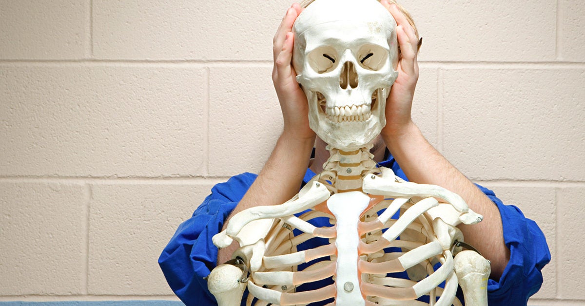
Back Human Bones Labeled : Labeled Skeleton Back View Of Male Skeleton Male Skeleton Anatomy Human Anatomy / The cervical spine is further divided into two parts;. The back comprises the spine and spinal nerves, as well as several different muscle the spine is composed of 33 bones called vertebrae, which stack together to form the spinal canal. Muscle anatomy triceps 12 photos of the muscle anatomy triceps. C1 is termed the atlas and c2 the axis. Below the lumbar spine is a bone called the sacrum, which makes up the back part of the pelvis. Human backbone diagram, bone, human backbone diagram.
Overview of bones & the axial skeleton. The labeled human skeleton system is comprised of 206 different bones of various sizes and shapes, all with the primary purpose of providing support, protection, and shape to the human body. Your skeleton can be divided into two main parts. The back comprises the spine and spinal nerves, as well as several different muscle the spine is composed of 33 bones called vertebrae, which stack together to form the spinal canal. Muscle anatomy triceps 12 photos of the muscle anatomy triceps.
Bone And Skeleton Fun Facts For Kids from easyscienceforkids.com See human skull anatomy stock video clips. Bone structure diagram human foot 12 photos of the bone structure diagram human foot bone structure diagram human foot, bone, bone structure diagram human foot. This bone rests between the scaphoid and triquetrum in the proximal row, near the. The back comprises the spine and spinal nerves, as well as several different muscle the spine is composed of 33 bones called vertebrae, which stack together to form the spinal canal. The ethmoid bone is one of the 8 bones of the cranium. Female muscle groups anatomical fitness vector illustration, sports training informative chart. Human backbone diagram, bone, human backbone diagram. Sciatica medical health care vector illustration scheme with lower spine and sciatic nerve pain in leg.
Below the lumbar spine is a bone called the sacrum, which makes up the back part of the pelvis.
Some individuals may also have additional (i.e., supernumerary) cervical ribs or lumbar vertebrae. When you look at the skeleton from behind, you can clearly see the spine running down the back, and the broad plates of the shoulder blades and pelvis. Using this atlas of human anatomy of the spine and back. The muscles of the back can be arranged into 3 categories based on their location: There is only one movable joint in the skull. Your skeleton can be divided into two main parts. Human anatomy and physiology lab (bsb 141) module 6: Superficial back muscles, intermediate back muscles and intrinsic back muscles.the intrinsic muscles are named as such because their embryological development begins in the back, oppose to the superficial and intermediate back muscles which develop elsewhere and are therefore classed as extrinsic muscles. While many of us take the benefits of a healthy spine for granted, spinal pain is a sharp reminder of how much we depend on our back in daily life. Sciatica medical health care vector illustration scheme with lower spine and sciatic nerve pain in leg. Facet joints connect each vertebra, with fluid supporting. Backbone diagram with vertebrae, disks and nerves. It comprises of a series of bones called the vertebrae of varying sizes extending from the skull to the small of the back.
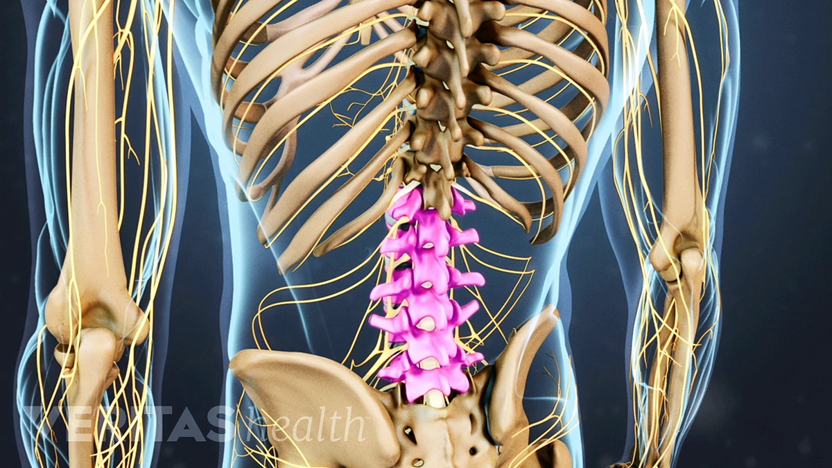
These bones work together to provide flexibility to the trunk, support the muscles of the trunk, and protect the spinal cord and spinal nerves of the back. C1 is termed the atlas and c2 the axis. It is situated towards the dorsal part of the torso. Nerves in your lower back five pairs of lumbar spinal nerves labeled l1 to l5 branch off your spinal cord and exit through small holes between the vertebrae. The lumbar spine is the lower back that begins below the last thoracic vertebra (t12) and ends at the top of the sacral spine, or sacrum (s1).
Flat Bones Definition Examples Diagram And Structure from post.healthline.com The lumbar spine is the lower back that begins below the last thoracic vertebra (t12) and ends at the top of the sacral spine, or sacrum (s1). Vertebrae separated by intervertebral discs. The spine is composed of 33 bones called vertebrae, which stack together to form the spinal canal. The vertebral column is the defining characteristic of a vertebrate in which the notochord (a flexible rod of uniform composition) found in all chordates has been replaced by a segmented series of bone: This bone is on the thumb side of the hand near the radius.; The cervical spine is further divided into two parts; Flexibility especially in the lower back and neck allowing us to bend and twist in a full variety of movements strength provided by the bones discs joints and supportive muscles and connective tissue that allows us to stand upright and move about with precision. This protects the spinal cord inside.
Vertebrae, bones, joints, ligaments, muscles, muscular system, fascia, arteries, veins, nerves and various adjacent organs.
While many of us take the benefits of a healthy spine for granted, spinal pain is a sharp reminder of how much we depend on our back in daily life. Superficial back muscles, intermediate back muscles and intrinsic back muscles.the intrinsic muscles are named as such because their embryological development begins in the back, oppose to the superficial and intermediate back muscles which develop elsewhere and are therefore classed as extrinsic muscles. Nerves in your lower back five pairs of lumbar spinal nerves labeled l1 to l5 branch off your spinal cord and exit through small holes between the vertebrae. These bones work together to provide flexibility to the trunk, support the muscles of the trunk, and protect the spinal cord and spinal nerves of the back. The vertebral column of the lower back includes the five lumbar vertebrae, the sacrum, and the coccyx. The back comprises the spine and spinal nerves, as well as several different muscle the spine is composed of 33 bones called vertebrae, which stack together to form the spinal canal. The eight bones of the wrist are:. Below the lumbar spine is a bone called the sacrum, which makes up the back part of the pelvis. The most common variations include sutural (wormian) bones, which are located along the sutural lines on the back of the skull, and sesamoid bones which develop within some tendons, mainly in the hands and feet. Vertebrae, bones, joints, ligaments, muscles, muscular system, fascia, arteries, veins, nerves and various adjacent organs. Human back bone chart, find out more about human back bone chart. This bone is on the thumb side of the hand near the radius.; The muscles of the back can be arranged into 3 categories based on their location:
The lumbar spine is the lower back that begins below the last thoracic vertebra (t12) and ends at the top of the sacral spine, or sacrum (s1). See sacrum (sacral region) the sacrum is connected to part of the pelvis (the iliac bones) by the sacroiliac joints. The eight bones of the wrist are:. It comprises of a series of bones called the vertebrae of varying sizes extending from the skull to the small of the back. Overview of bones & the axial skeleton.
Understanding Lower Back Anatomy from embed.widencdn.net 12 photos of the human back bone chart. This bone is shaped like a triangle that fits between the two halves of the pelvis, connecting the spine to the lower half of the body. Posted on august 7, 2015 by admin. The vertebral column of the lower back includes the five lumbar vertebrae, the sacrum, and the coccyx. It is situated towards the dorsal part of the torso. This bone rests between the scaphoid and triquetrum in the proximal row, near the. The labeled human skeleton system is comprised of 206 different bones of various sizes and shapes, all with the primary purpose of providing support, protection, and shape to the human body. The spine's four sections, from top to bottom, are the cervical (neck), thoracic (abdomen,) lumbar (lower back), and sacral (toward tailbone).
To learn all about the skeleton system in the human body, check out this guide.
This diagram depicts skeletal images 744×1314 with parts and labels. 12 photos of the human back bone chart. The bones of the skull. This bone is shaped like a triangle that fits between the two halves of the pelvis, connecting the spine to the lower half of the body. C1 is termed the atlas and c2 the axis. Muscle anatomy triceps 12 photos of the muscle anatomy triceps. To learn all about the skeleton system in the human body, check out this guide. Back anatomy stock photos and images. The part of the nerve that emerges out of the spine is called the nerve root. It comprises of a series of bones called the vertebrae of varying sizes extending from the skull to the small of the back. Human back bone chart, find out more about human back bone chart. The muscles of the back can be arranged into 3 categories based on their location: It runs down the centre of the body.
The human backbone is also known as the vertebral column or the spinal column human back bones. Human organ diagram body organs diagram human body muscles human body organs anatomy organs anatomy bones heart anatomy body muscle anatomy human body anatomy.